|
|
LATEST NEWS
NEW PUBLICATIONS

Portal vein thrombosis: diagnosis, management, and endpoints for future clinical studies
Elkrief L. et al - The Lancet Gastroenterology & Hepatology - 2024
Elkrief L. et al - The Lancet Gastroenterology & Hepatology - 2024

Performance of spleen stiffness measurement to rule out high-risk varices in patients with porto-sinusoidal vascular disorder
Moga L. et al - Hepatology - 2024
Moga L. et al - Hepatology - 2024
DECISION : FIRST PUBLICATION

Sara Palomino-Echeverria et al. - Journal of Translational Medicine - 2024
New tool identifies patient subgroups for personalized management of acutely decompensated cirrhosis
ClustALL, a newly developed stratification method provides insights in understanding differences among patients with acutely decompensated cirrhosis showing potential for clinical applications.
Findings from a recent study, published on 27 June 2024 in Journal of Translational Medicine, highlight the robustness of ClustALL, a novel computational tool, in identifying differences among patients with acutely decompensated cirrhosis, which could lead to more personalized and effective healthcare.
NEW PUBLICATION

New publication of the team :
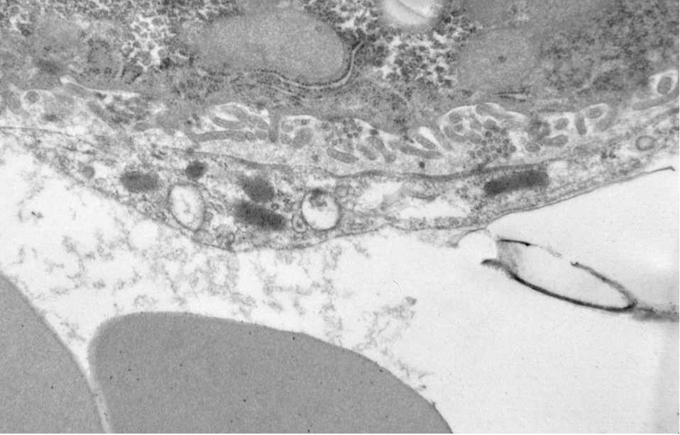
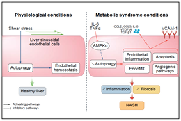
Loss of fenestrae in liver sinusoidal endothelial cells contributes to MASLD
Nadia Ciriaci, Pierre-Emmanuel Rautou & Johanne Poisson
Nature Cardiovascular Research - June 2024
NEW GRANT

Prof. Pierre-Emmanuel Rautou has obtained new funding: COST Action.
The COST (Cooperation in Science and Technology) program is a European funding initiative aimed at strengthening collaboration among researchers and scientists in Europe. COST Actions are cooperative projects that bring together researchers from various countries to work on specific topics over a period of four years.
The COST (Cooperation in Science and Technology) program is a European funding initiative aimed at strengthening collaboration among researchers and scientists in Europe. COST Actions are cooperative projects that bring together researchers from various countries to work on specific topics over a period of four years.
BRAINSTORMING 2024
TEAM RAUTOU
EASL CONGRESS 2024
5-8 june MILAN

An event that brings together the global hepatology community, happening from June 5-8, 2024, in Milan, Italy, and online.
The EASL Congress, Europe's largest event in hepatology, will host over 7,000 professionals, including clinicians, researchers, allied health professionals, patient representatives, and industry experts.
This four-day congress will feature interactive and hands-on learning sessions from renowned faculty, offering the liver community unparalleled opportunities to present their research, network, and engage directly with top experts in hepatology.

|

|

Prof. Pierre-Emmanuel Rautou organized a Postgraduate Course with Prof. Cristina Ripoll and Prof. Ton Lisman
The role of vascular biology in chronic liver disease : implications for clinical management.
Two team members gave lectures at this occasion:
- Medical management of portal vein thrombosis in patients without cirrhosis by Dr Aurélie Plessier
- Coagulation changes in patients with cirrhosis : Case 2 : Bleeding after dental extraction by Prof. Laure Elkrief
|
Pr. Pierre Emmanuel Rautou presented the EASL clinical practice guidelines on vascular liver diseases |

|

|
Dr Johanne Poisson presented a Workshop: My biggest mistakes and what I learned from them When mistakes lead to new discoveries |
|
Dr Shantha Valainathan presented his work on biomarker and acute decompesation |

|
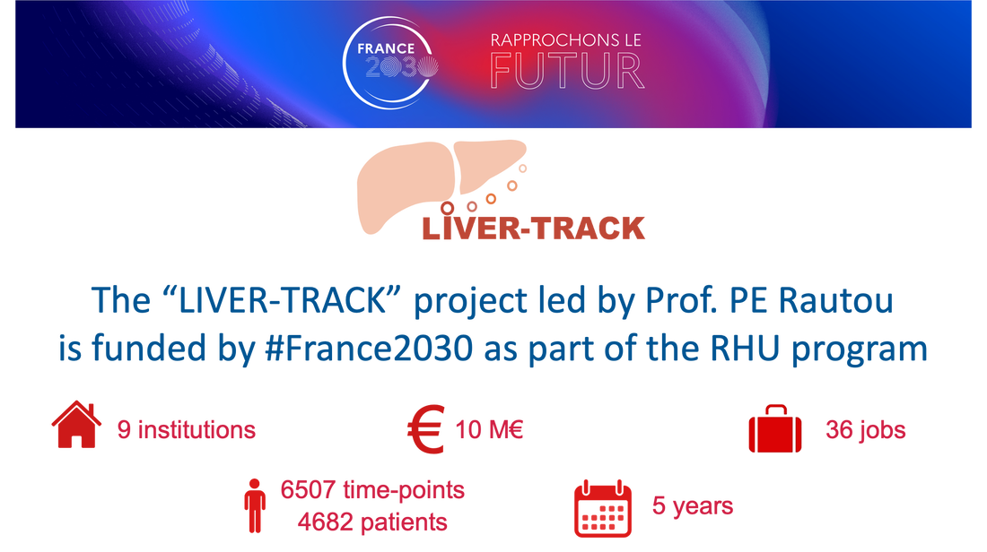
KICK-OFF MEETING LIVER TRACK
Liver Track ?
The objective of LIVER-TRACK is to address two unmet medical needs in patients with cirrhosis:
1) the estimation of the risk of decompensation of cirrhosis, and 2) the estimation of the risk of liver cancer development.
By bringing together the highly innovative measurements of circulating extracellular vesicles (EVs), an untapped source of biomarkers in liver diseases, with state-of-the-art existing biomarkers including FibroTest, cancer proteins and SNPs, LIVER-TRACK will create a Test for Decompensation and a Test for Liver Cancer.
These tests will be turnkey solutions to predict the evolution of patients with cirrhosis in routine clinical practice.
https://www.liver-track.com/
The liver Track kick-off took place on 23 May.
NEW PUBLICATION

Prognosis algorithms for acute decompensation of cirrhosis and ACLF
S.R. Valainathan et al.
Liver international - 26 march 2024
S.R. Valainathan et al.
Liver international - 26 march 2024
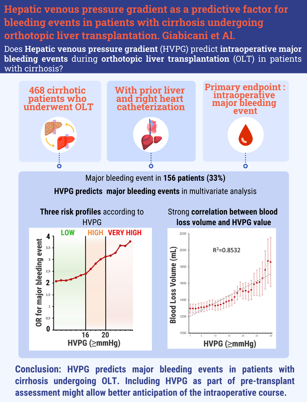
Predictive Role of Hepatic Venous Pressure Gradient in Bleeding Events Among Cirrhotic Patients Undergoing Orthotopic Liver Transplantation
M. Giabicani
YI Afterwork Series Webinar – Navigating through an academic career: Step-by-step
With Dr. Johanne Poisson
Advancing in a scientific career feels more like a marathon than a sprint. In addition to the academic challenges, life often gives you lemons. This webinar focuses on the most relevant steps throughout a research career and the skills needed to succeed:
- How to plan the foreseeable and cope with the unexpected changes.
- How to turn obstacles into opportunities for personal growth.
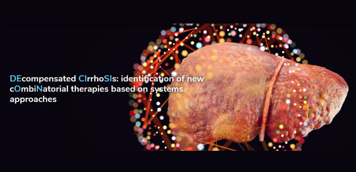
DECISION Project: Advancing Treatment Strategies for Decompensated Cirrhosis
The objective of the DECISION project is to enhance the understanding of the pathophysiology of decompensation of #cirrhosis leading to acute-on-chronic liver failure (#ACLF) or death. This consortium will take advantage of already existing large and clinically well-characterized cohorts to ultimately develop prognostic and response tests and combinatorial therapies tailored to the needs of individual patients to decrease the risk of short-term death.
Follow DECISION
🌐 Visit the DECISION website: https://decision-for-liver.eu/
🤳 Follow DECISION on social media: Twitter / X: / decision4liver LinkedIn: / decision-project
This project has received funding from the European Union’s Horizon 2020 research and innovation programme under grant agreement No 847949.
This video reflects only the presenter's view and the European Commission is not responsible for any use that may be made of the information it contains.
Video production & subtitles: Department of Medicine, University of Padova, Padova, Italy
© 2024 DECISION
NEW PUBLICATION
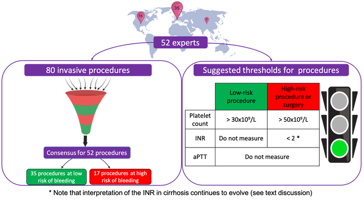
Expert opinion on bleeding risk from invasive procedures in cirrhosis
Alix Riescher-Tuczkiewicz et al.
JHEP Reports - Dec 2023
Alix Riescher-Tuczkiewicz et al.
JHEP Reports - Dec 2023
DECISION 5th GA Meeting - Padova, Italy
Members of the DECISION Consortium met in Padova, Italy, to discuss recent research results in preparation of several manuscripts shedding some new light on the pathophysiology of acute decompensation of cirrhosis and development of acute-on-chronic liver failure (ACLF) – a syndrome associated with high short-term mortality. After three years of intensive work, the upcoming COMBAT trial will use newly identified biomarkers to predict disease progression and test safety and effectiveness of a combinatorial drug therapy.
https://decision-for-liver.eu/
https://decision-for-liver.eu/
BRAINSTORMING TEAM RAUTOU 2023
Between research and walk in the Baie de Somme
Journée recherche FILFOIE 2023
Actualité sur les maladies vasculaires du foie
Pierre-Emmanuel RAUTOU
LAST PUBLICATIONS
|
Endothelial autophagy is not required for liver regeneration after partial hepatectomy in mice with fatty liver Hammoutene A. et al. Liver International. June 2023 |
Journée recherche FILFOIE 2023
Actualité sur les maladies vasculaires du foie
Pierre-Emmanuel Rautou
LAST PUBLICATIONS
|
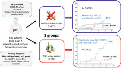
Hepatocyte-derived biomarkers predict liver-related events at 2 years in Child-Pugh class A alcohol-related cirrhosis. Elkrief L, et al. J Hepatol. 2023 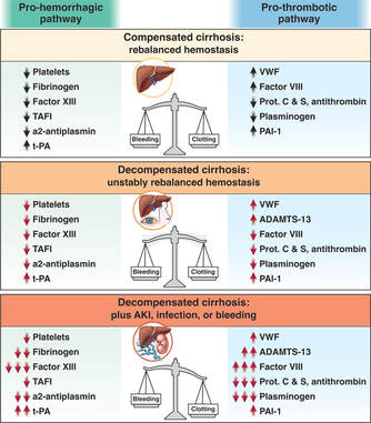
|
Bleeding and Thrombotic Complications in Patients with Cirrhosis: A State of the Art Appraisal. Rautou PE, Caldwell SH, Villa E. Clin Gastroenterol Hepatol. 2023 |
EASL STUDIO
EASL-EF CLIF and EU Grants: Exploring novel mechanism and treatment of chronic liver failure
Through widespread multidisciplinary collaboration, consortia and EU grants provide unique opportunities to address critical questions.
- What opportunities exist for early career scientist?
- Which elements are essential for success?
- How have these partnerships contributed to advancements in the field?
- Prof. Thomas Berg (Moderator)
- Prof. Rajiv Jalan (Moderator)
- Prof. Minneke Coenraad (Faculty)
- Dr Cornelius Engelmann (Faculty)
- Prof. Pierre-Emmanuel Rautou (Faculty)
LAST PUBLICATIONS
|
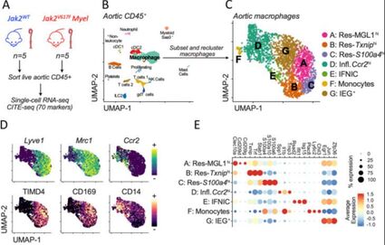
JAK2V617F mutation drives vascular resident macrophages toward a pathogenic phenotype and promotes dissecting aortic aneurysm Rida Al-Rifai et al. Nat Commun. 2022 Collaboration with Team Ait-Oufella |
|
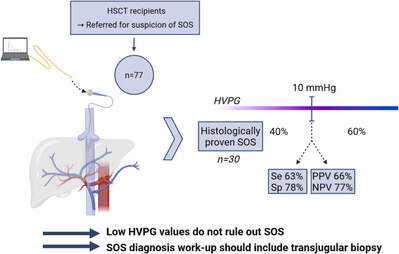
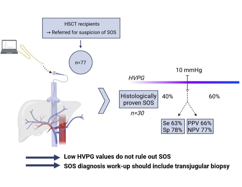
Hepatic venous pressure gradient in sinusoidal obstruction syndrome: diagnostic value and link with histological lesions Simon B Gressens. JHEP Rep. 2022 |
|
|
EASL STUDIO
YI Choice: how to build consortia and registries
The value of scientific collaboration and data sharing through registries and consortia is unequivocal. But what are the main challenges to building a consortium and how do we overcome them.
Join the faculty as they provide you with a practical, experience-based view of how to build a successful consortium.
Join the faculty as they provide you with a practical, experience-based view of how to build a successful consortium.
- Dr Marta Afonso (Moderator)
- Dr Mattias Mandorfer (Moderator)
- Prof. Jesús Bañales (Faculty)
- Prof. Rajiv Jalan (Faculty)
- Prof. Pierre Emmanuel Rautou (Faculty)
DECISION : 7th DECISION Steering Committee Meeting
End of March 2023, the DECISION project marked its 7 steering committee meeting in Pamplona. Two days packed with updates on the overall progress of the project, presented by work package leaders and project members. Lively discussions cemented the project’s path for the next months.
https://twitter.com/i/status/1643223082927022080
https://twitter.com/i/status/1643223082927022080
EASL STUDIO
YI Choice: Gender equality in academics — reducing the gap
In honour of International Women’s day, this episode of EASL Studio raised awareness about the gender gap in academic research and debating how to improve gender diversity.
- Dr Johanne Poisson (Moderator)
- Prof. Eric Trépo (Moderator)
- Prof. Dina Balabanova (Faculty)
- Dr Ea Høg Utoft (Faculty)
EASL STUDIO
CIRRHOSIS AND COMPLICATIONS
In this episode of EASL Studio the experts discuss a non-cirrhotic portal vein thrombosis (PVT):
Faculty:
Please click here to access the podcast version of this EASL Studio episode.
- What is PVT in the absence of cirrhosis,
- The role of interventional radiology in the management of portal cavernoma, and
- An update from the VALDIG portal Vein Thrombosis meeting
Faculty:
- Prof. Pierre-Emmanuel Rautou (Moderator)
- Prof. Juan-Carlos Garcia-Pagan (Faculty)
- Dr Aurélie Plessier (Faculty)
Please click here to access the podcast version of this EASL Studio episode.
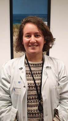
Congratulations to Louise Biquard for his PhD defense
DECISION
4th General Assembly (GA) meeting in Madrid
The 4th GA meeting of DECISION took place from 19-21 October 2022 in Madrid.
JAK2V617F mutation drives vascular resident macrophages toward a pathogenic phenotype and promotes dissecting aortic aneurysm
Rida Al-Rifai - Nature Communications - 2022
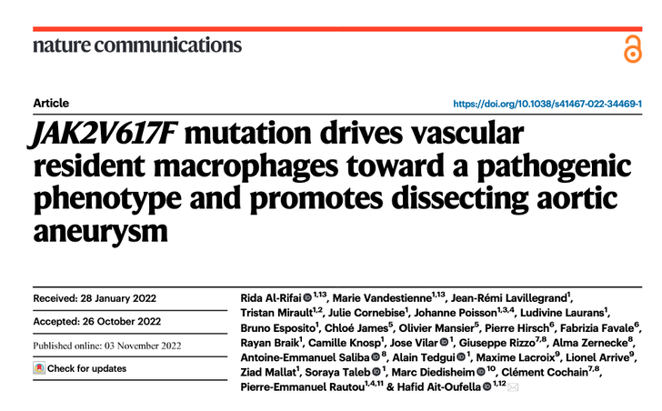
New paper from Hafid Ait-Oufella's team in collaboration with ou team
JAK2V617F mutation drives vascular resident macrophages toward a pathogenic pheno-type and promotes dissecting aortic aneurysm.
PUMS college= "How to take care of your liver"
with Prof. Pierre-Emmanuel Rautou, Dr. Audrey Payancé and Prof. Laurent Castéra
Predicting bleeding after liver biopsy using comprehensive clinical and laboratory investigations: a prospective analysis of 302 procedures
J. Bissonnette, A. Riescher-Tuczkiewicz, et al.
Journal of thrombosis and haemostasis. 2022
- Extensive hemostasis workup does not improve the prediction of liver biopsy-related bleeding
- Pain 2 hours after liver biopsy was associated with the occurrence of biopsy related-bleeding
Link
- Pain 2 hours after liver biopsy was associated with the occurrence of biopsy related-bleeding
Link
Hepatic venous pressure gradient in sinusoidal obstruction syndrome: diagnostic value and link with histological lesions
Simon B. Gressens, et al. J HEP REPORTS 2022
INTERNATIONAL LIVER CONGRESS
EASL 2022
EASL Studio : Teatime with Pr. Pierre-Emmanuel Rautou and Dr. johanne Poisson
|
Prof Pierre-Emmanuel Rautou moderates a studio session including Prof Pere Gines, Dr Mattias Mandorfer, Prof Debbie Shawcross, and Prof Dominiquer Thabut: great discussions! Link |
|
Dr Johanne Poisson has been invited as faculty in EASL studio for the ILC 2022 alongside with Prof. Helena Cortez-Pinto and Prof. Aleksander Krag. This session was moderated by Prof. Tobias Böttler and Assoc. Prof. Jean-Charles Nault. Link |
|
Picture EASL

Portal vein recanalisation alone to treat severe portal hypertension in non-cirrhotic patients with chronic extrahepatic portal vein obstruction
Artru F et al; JHEP Rep. 2022
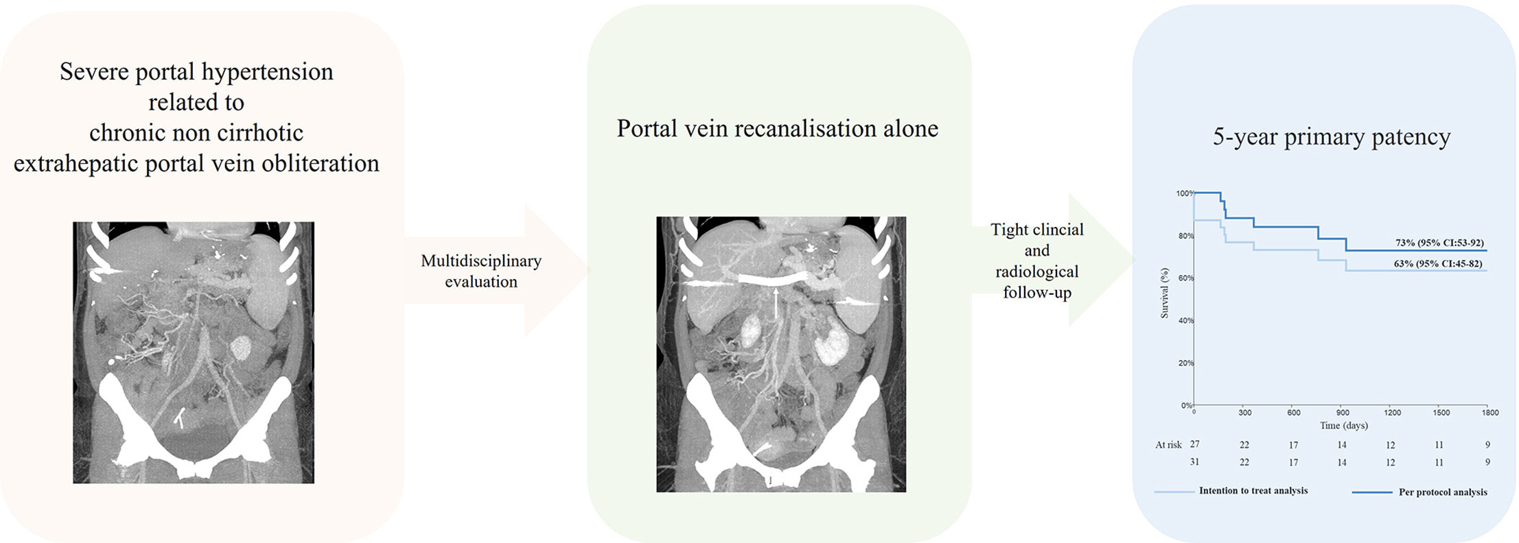
In patients with technical success, 5-year primary patency was 73%, and was associated with improved muscle mass and decreased spleen volume at 1 year.
Link
Link
Prof Pierre-Emmanuel Rautou interviews the chairs of European and American guidelines on coagulation in liver diseases about the interest of anticoagulants in cirrhosis to improve patients' outcome
Contrast-enhanced CT and liver surface nodularity for the diagnosis of porto-sinusoidal vascular disorder: a case-control study
Shantha Ram Valainathan et al. - Hepatology - 2022
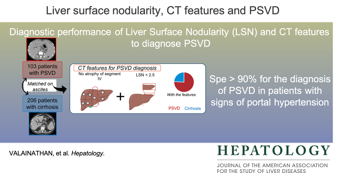
CT-scan to raise suspicion of Porto-Sinusoidal Vascular Disorder (PSVD):
- Liver Surface Nodularity and size of segment IV have a > 90% specificity for diagnosis of PSVD
- One more tool to help identify patients with this rare disease
A new video dedicated to patients, explaining portosinusoidal vascular disorders
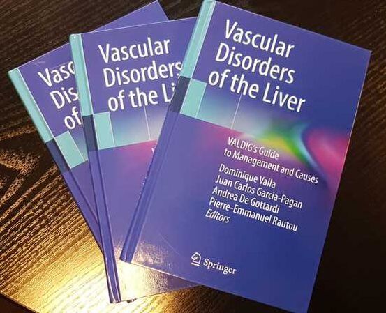
Vascular Disorders of the Liver
|
VALDIG's Guide to Management and Causes
Link |
|
|
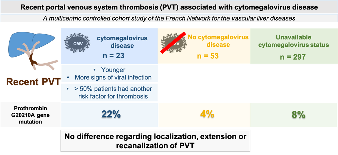
Multicenter study on recent portal venous system thrombosis associated with cytomegalovirus disease
Chloé De Broucker et al. - J. Hepatol - 2021
CMV disease triggers for portal vein thrombosis : => a special link with factor II gene mutation => do not forget to screen for all risk factors for thrombosis => no impact on recanalisation. For more details, please read our paper |
EASL TAKEWAYS - Cirrhosis and complications
Discover cirrhosis and complications with Professor Didier Samuel, Professor Andrés Cárdenas and Professor Pierre-Emmanuel Rautou
ERN Rare-Liver on-demand Webinar: Porto sinusoidal vascular disease
Podcast of Prof PE Rautou on porto-sinusoidal vascular disease : link
The International Liver Congress - EASL STUDIO
Prof U Beuers interviews Prof PE Rautou on porto-sinusoidal vascular disease
|
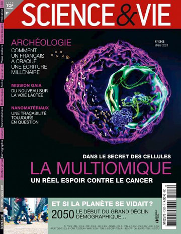
In the March 2021 issue of "Sciences et vie", multiomics are described in great detail. At this occasion, Prof. Pierre-Emmanuel Rautou explains the approach developed in the European project he coordinates, using omics to improve diagnosis and prognosis of patients with liver diseases : “C’est pourquoi nous avons lancé avec 20 autres centre européens un vaste projet d’analyse multiomique sur une cohort qui compte déjà 2200 patients.” |
Pierre-Emmanuel Rautou and Valerie Paradis obtained an ARC funding for the project "Endothelial autophagy in liver carcinogenesis related to NonAlcoholic Fatty Liver Disease : Role and potential therapeutic target"
|
|
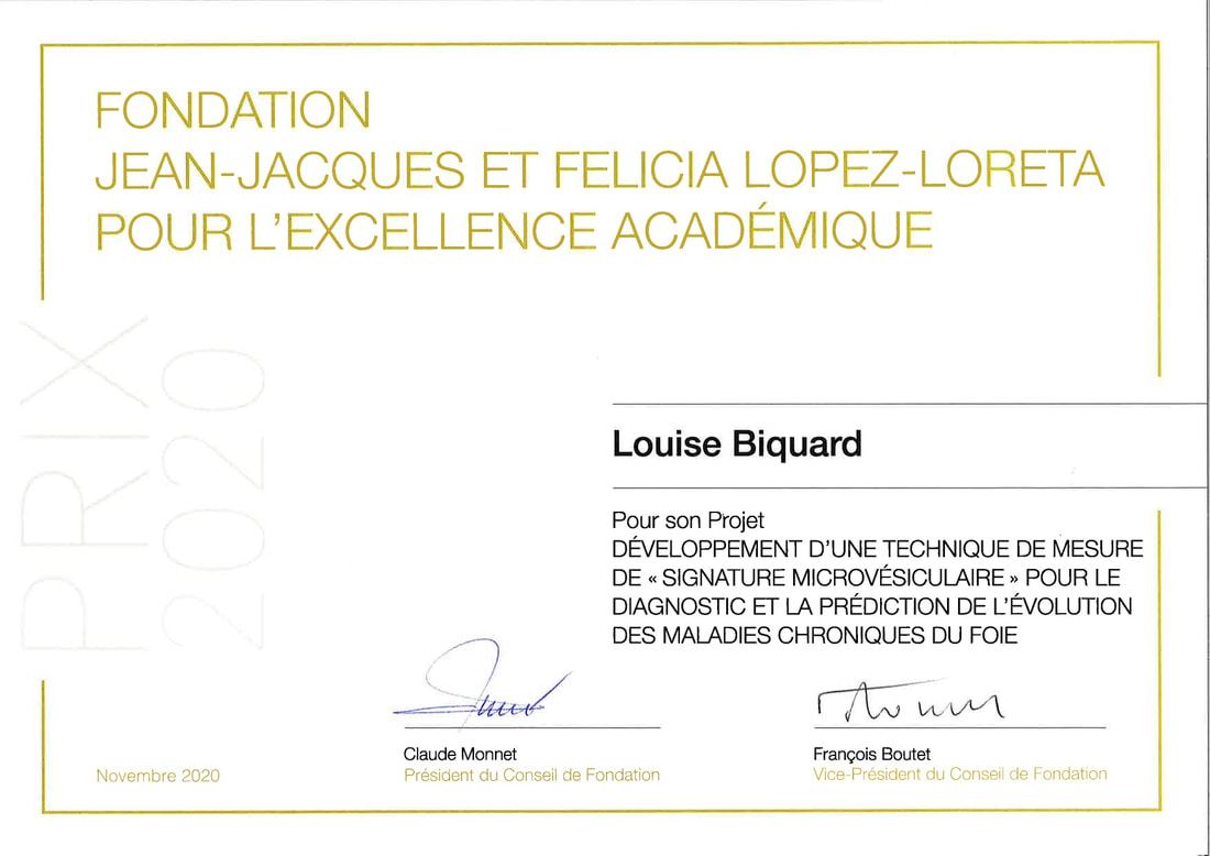
Congratulations to Louise Biquard for her great award
EASL Digital ILC 2020 Takeaways - Cirrhosis and complications
The DECISION project coordinated by Prof Rautou
European researchers seek to reduce the number of patients dying from cirrhosis
Visite the website for more informations => here
European researchers seek to reduce the number of patients dying from cirrhosis
Visite the website for more informations => here
|
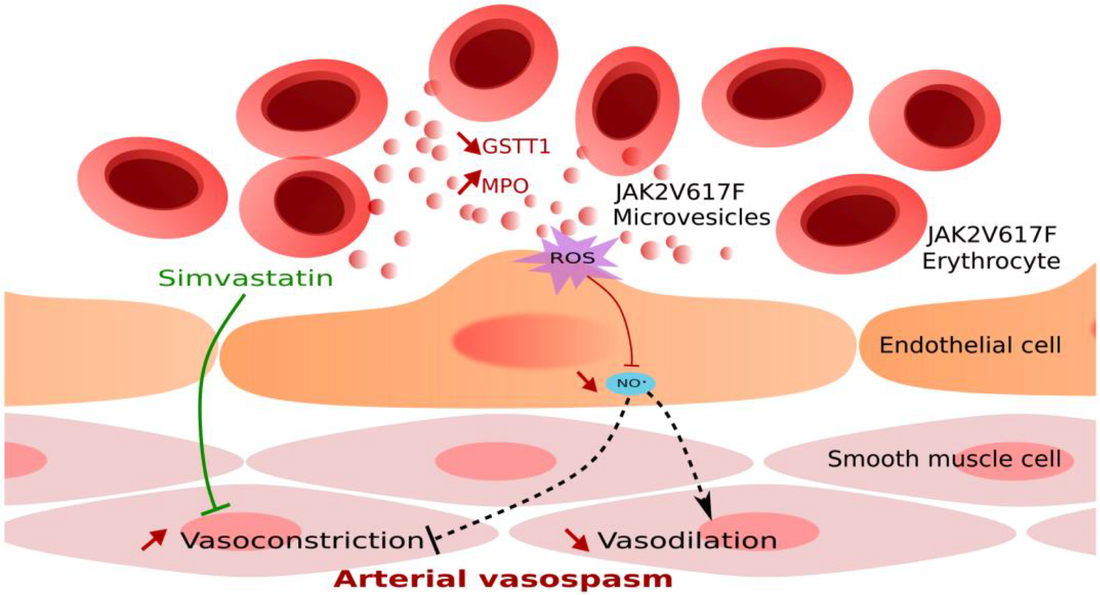
New Publication
Erythrocyte-derived microvesicles induce arterial spasms in JAK2V617F myeloproliferative neoplasm |
|
Our lab received the "EMERGENCE grant" by the city of Paris for the project “Predicting Outcome of patients with cirrhosis using circulating extracellular Vesicles”
|
Pierre-Emmanuel RAUTOU obtained the ATIP AVENIR funding which allows young scientists to create and lead a team within an established Inserm laboratory in France in January 2019
|
SPONSORS
|
|
|
|
|
|
|

|

|
|
|
|

|
Centre de recherche sur l'inflammation
UMR 1149 Inserm - Université de Paris
ERL CNRS 8252
Faculté de médecine site Bichat
16 rue Henri Huchard F-75018 Paris
UMR 1149 Inserm - Université de Paris
ERL CNRS 8252
Faculté de médecine site Bichat
16 rue Henri Huchard F-75018 Paris